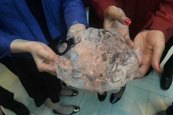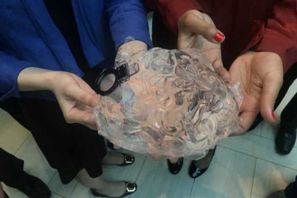
Bengaluru : A 36-year-old patient suffering from motor area tumor successfully underwent Awake brain surgery at Fortis Hospital, Bannerghatta Road. The team of doctors led by Dr Satish Satyanarayana – Additional Director- Neuro Surgery along with Dr Simha-Senior Anaesthetist at Fortis Hospital, Bannerghatta Road.
Awake Brain Surgery or Awake Craniotomy is a surgical technique that enables surgeons to avoid injury to critical regions of the brain during surgery and are helpful in cases where cortical mapping or continuous monitoring of neurological functions are expected to improve outcomes.
The patient was presented with a history of focal seizure of right upper and lower limbs a year ago followed by a second episode of seizure two months later. He started witnessing recurrent seizures coupled with loss of consciousness for about 5minutes. Post thorough diagnosis at Fortis Hospitals, it was found that the size of lesion has increased in the brain, hence he was advised surgical management.
Dr Satish Satyanarayana – Additional Director- Neuro Surgery at Fortis Hospital, Bannerghatta Road, explains, “The size of the tumor was found to have 2.14*2.51*1.79cm with perilesional edema, therefore we recommended him to undergo neuro navigation guided left posterior frontal navigation guided, awake craniotomy with neuro monitoring for micro neurosurgical excision of the intrinsic brain tumor in motor cortex by 3D Zeiss operative microscope and intraop fluoresein imaging for safe surgery.
Normally, brain tumors are operated on patients under general anaesthesia which facilitates patients not be aware and cooperate for the procedure. However, for certain tumors in or near vicinity of vital brain regions like Motor or speech area, the safety of surgery lies in patient being able to be monitored in real time by the neuro anaesthesiologist Dr Simha-Senior Anaesthetist. We used cutting edge other supports like 3D neuronavigation mapping out and avoided major pathways vital for functioning as well as intraop tumor fluorescence which highlighted tumor and differentiated it from neural structures. With the help of this technique, the surgery became extremely safe and we had a perfect neurological outcome.
Initially, the patient was very apprehensive, however, after several rounds of counselling and briefing session with doctors, he agreed to undergo surgery and was very cooperative during the procedure. Thus we were able to perform all the required tasks perfectly and ended up with zero deficits despite tumor being located exactly in the motor strip.”, he added.

